Cyclisation is one of the most dependable ways to make a peptide behave more like a drug, tightening its fold, sharpening target engagement, and slowing proteolysis. Yet most cyclisation chemistries run in a test tube, after the peptide has already been made and purified. Executing the same coordination chemistry inside a living cell is far harder, because the cytoplasm is crowded with competing thiols, reductants, and metal-binding molecules that readily derail a metal-thiolate reaction. Bismuth, a compact and highly thiophilic single-atom scaffold, offered an appealing route. The peptide-bismuth bicycle chemistry pioneered by Christoph Nitsche and coworkers had already shown that a lone Bi(III) centre can staple cysteine-rich peptides into stable bicycles, first in solution, in a paper first-authored by Saan Voss, and later encoded on phage in work led by Sven Ullrich, all three of whom have been featured on our site. Even so, that chemistry had stayed outside the cell, its crowded reducing interior assumed to be hostile territory.
Researchers in the Mason Group at the University of Bath, publishing in Communications Chemistry, have now, for the very first time, shown that this particularly compact cyclisation chemistry, bismuth(III)-thiolate coordination, can be carried out inside living E. coli during recombinant expression. The approach is disarmingly simple. Supplement the growth medium with a bismuth salt, and the metal enters the cytoplasm and chemoselectively clamps onto cysteine residues in the expressed peptide, closing the ring in situ. Of the two salts tested, BiK3[citrate]2 was better tolerated than BiBr3, cyclising a three-cysteine test peptide at roughly 96 percent efficiency at 250 µM while leaving the bacteria healthy.
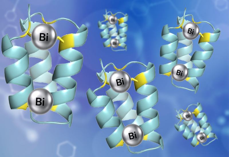
The team then pushed the chemistry beyond single loops. Starting from a computationally designed three-helix bundle, they engineered a miniprotein carrying two spatially separated cysteine triads. Inside the cell, this scaffold picked up two bismuth atoms to form a tetracyclic miniprotein, a level of intracellular structural reinforcement not previously accessible. The payoff was substantial. Fractional helicity roughly doubled, from 29 percent in the linear form to 62 percent once constrained, the fold held to around 80 °C, and serum half-life climbed to 361 minutes. Because bismuth(III)-thiolate coordination is remarkably inert, the constraint survived even strong reducing agents such as TCEP, which readily unravel ordinary disulfides.
The real test was whether this chemistry could be wired into a functional screen. The group coupled intracellular bismuth cyclisation to an alpha-synuclein protein-fragment complementation assay, screening a 16.8-million-member bicyclic library directly under selective, bismuth-cyclising conditions with better than 99.9 percent coverage. Iterative enrichment converged on a single sequence, αS-BiP1, that reduced alpha-synuclein aggregation in a concentration-dependent manner. Tellingly, the linear version of the same peptide was far weaker, confirming that the bismuth-locked bicyclic shape, not the sequence alone, drives the activity. Alpha-synuclein aggregation sits at the heart of Parkinson's disease, making it a fitting proof of principle.
The elegance here lies in timing. Because the peptide is constrained as it is expressed, its sequence and its shape are optimised under the same selective pressure that defines function, keeping genotype and phenotype tightly linked. That sets the method apart from post-expression and display-based strategies, where the constraint is imposed outside the selective environment. Building on the group's earlier work using bis-alkylating reagents inside cells, bismuth coordination now adds a compact, single-atom route to multicyclic architectures. It widens the range of peptide and miniprotein topologies that live-cell platforms can discover, and points toward constrained intracellular ligands with real therapeutic potential.